Hip Anatomy
Hip Anatomy
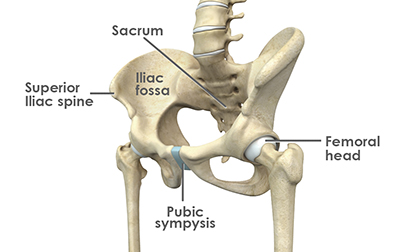
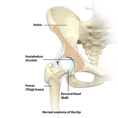
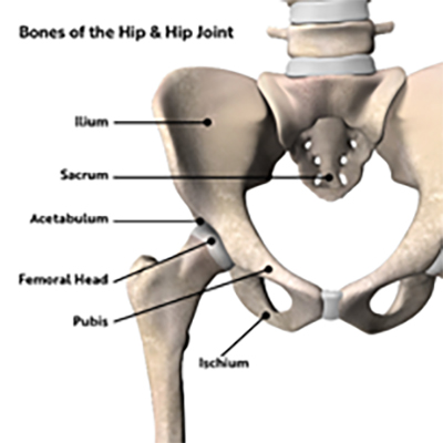
The hip joint is a “ball and socket” joint. The “ball” is known as the femoral head and the “socket” is the part of the pelvis known as the acetabulum. Both the femoral head and the acetabulum are coated with articular cartilage. Like all joints, the hip has synovial or joint fluid, acting as a lubricant, which allows for smooth, painless movement within the hip joint.
The hip, like the shoulder, has a labrum, a ring of rubbery cartilage around the rim of the acetabulum, which deepens the hip socket and acts as the suction seal of the hip joint. The intact labrum seals the lubricating fluid within the hip and contributes to stability of the joint.
One of the most common causes of hip pain involves damage to the labrum, described most often as a labral tear. When the labrum is torn, the “suction seal” is broken, and the joint may lose its stability and lubrication. This can progress to loss of cartilage, or even arthritis. Arthritis is the damage to the articular cartilage of the joint that lines on the head of the femur and the acetabulum, or socket.
The joint capsule is an envelope of ligaments that encloses the hip, and is also essential for stability. The hip also contains a ligament known as the ligamentum teres, which connects the femoral head to the acetabulum. Both the capsule and the ligamentum teres can be injured in an unstable hip.
Diagnosis of Hip Problems
- Diagnosis of hip injuries can be complex. Studies have shown that as many as 60% of patients requiring a hip arthroscopy were initially misdiagnosed. Hip pain may mimic many other conditions, including sciatica, spinal problems, muscle strains, and sports hernias.
- Obtaining the correct diagnosis is of paramount importance in directing appropriate treatment. A systematic approach to diagnosis may include several steps:
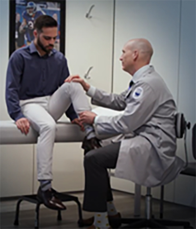
- Specialized clinical exam by a hip specialist
- Specific new X-ray views designed for viewing hip anatomy
- Specialized MRI scans at a center specializing in pre-arthritic and arthritic hip conditions.
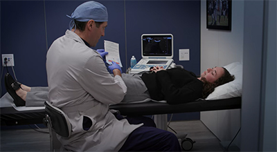
- Ultrasound guided Diagnostic intra-articular hip injections can help confirm the diagnosis and intra-articular involvement
- It is important to note that many patients are misdiagnosed despite having a physical exam, X-rays, and an MRI, because the studies were not the specific studies designed for pre-arthritic or early arthritic hip injuries.
- The providers at the American Hip Institute have been trained regarding the highest excellence in the hip physical exam, imaging techniques and performing diagnostic injections under ultrasound guidance.